
5. Cell Structure And Organization -

Part 01 - Cell,Totipotency and modern cell theory
Cell :


Totipotency :
Know the scientists
Kinds of cells :
A. Prokaryotic cells :
- Cell is called a structural and functional unit of life of all living organisms capable of independent existence and can perform all functions of life.

- Cells may be spherical, rectangular, flattened, polygonal, oval, triangular, conical, columnar, etc.
- Cell size varies greatly in various plants and animals. Some of them are not visible to naked eye. Some are barely visible while some are macroscopic.
- The smallest cell size can be seen in mycoplasma (0.3 μm in length), bacterial cell size is 3 to 5 μm, while the largest size of cell is seen in Ostrich egg (nearly 15cms).
- Longest cells are nerve cells.
- Cell theory was proposed by Schwann and Schleiden. However, in this theory, there was no explanation about formation of new cells.
- It was Rudolf Virchow (1855) who explained for the first time that new cells are formed by cell division from pre existing cells (Omnis cellula-e- cellulla).
- In later years, advanced research in cytology led to modification in cell theory, which is now known as Modern Cell Theory.
Totipotency :
- It is the capacity or the potential of living nucleated cell to differentiate and divide to form any other type of cell and thereby a complete new organism.
- A cell is totipotent because it has the entire genetic information of the organism in its nucleus.
- Embryonic animal cells are totipotent and termed as stem cells.
- Stem cells have great medical applications including cure for diseases.
Know the scientists
- A German botanist Matthias Schleiden (1838) examined number of plants and concluded that various tissues of plants are composed of different types cells.
- At that time, a British zoologist Theodore Schwann (1839) proposed that cells are bound by a thin membrane.
- He also explained about existence of cell wall as a unique character of plant cell.
- On the basis of his observation, he proposed that animals and plants are made up of cells and products of cells.
- All living organisms are made up of cells.
- Living cells arise from pre-existing cells.
- A cell is the structural and functional unit of life.
- Total activities of cells are responsible for activity of an organism.
- Cells show transformation of energy.
- Cells contain nucleic acids; DNA and RNA in the nucleus and cytoplasm.
Part 02 - Prokaryotic cells
Kinds of cells :
- Living organisms are grouped into two main categories -
- Prokaryotes and
- Eukaryotes.
- The prokaryotes have simple cellular organization while eukaryotes exhibit high degree of organization.
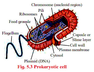
A. Prokaryotic cells :
- It has chemically complex protective cell envelop.
- It does not have well-defined nucleus and other membrane bound cell organelles.

Cell envelop is a three-layered structure with-
1.outer glycocalyx
2.middle cell wall and
3.inner plasma membrane.
Always remember :
Eukaryotic Cells :
Components of Eukaryotic cell :


3.suberin (endodermal cells of root)
4.wax
Middle lamella :
Primary wall :
Secondary wall :

Cell membrane / Plasma membrane/ biomembrane :
Fluid mosaic model :
 It is most accepted model of cell membrane.
Cytoplasm :
Endoplasmic Reticulum (ER):
Rough ER :
Golgi complex / Golgi apparatus / Golgi body:

always remember :
3.inner plasma membrane.
- Glycocalyx is present as either slime layer (loose sheath) or capsule (tough).
- Bacteria are better observed when stained.
- The most followed staining method is ‘Gram staining’ developed by Danish bacteriologist Hans Christian Gram.
- The cell wall is made up of peptidoglycan (in Gram positive bacteria) and murein (in Gram negative bacteria). It gives mechanical strength to the cell.
- Cell membrane is a phospholipid bilayer. All these structures give protection to the cell and also help in inter-cellular transport.
- In motile bacteria either cilia or flagella are found. Both are driven by rotatory movement produced by basal body (which works as motor).
- Other parts are filament and hook.
- Some other surface projections are the tubular pili (which help in inter-cellular communication) and fimbriae (for clinging to support).
- The cell membrane shows infoldings called mesosomes, which help in cell wall formation and DNA replication.
- Some bacteria especially photosynthetic cyanobacteria show more longer extensions called chromatophores.They carry photosynthetic pigments.
- The cytoplasm contains dense particles called ribosomes helping in protein synthesis.
- Ribosomes are described by their sedimentation rate in Svedberg units.
- Bacterial ribosome are 70S (composed of a larger subunit 50S + smaller subunit 30S).
- The term cell was first used by Robert Hooke (1665) in his book ‘‘Micrographic’’.
- Purkinje and Mohl (1835-37) discovered protoplasm.
- Camillo Golgi (1838) discovered the Golgi apparatus.
- Robert Brown (1881) discovered the Nucleus.
- Balbiani (1881) discovered chromosomes in salivary glands of Chironomus larva.
- Flemming (1882) studied cell division in detail and coined the term Mitosis.
- Porter (1945) discovered Endoplasmic Reticulum.
- C.Benda gave the name Mitochondria.
- C. de Duve (1955) discovered Lysosomes
Always remember :
- Genetic material in bacterium is a single chromosome made up of circular and coiled DNA. It remains attached to mesosome.
- This DNA undergoes a very typical replication pattern called as theta model of replication.
- The DNA is not associated with histone proteins (as in eukaryotes) hence not referred to as chromatin.
- Besides chromosomal DNA many bacteria show plasmids which are small circular DNA molecules carrying few genes.
- They are termed as extrachromosomal selfreplicating DNA molecules.
- They are of two basic types
- F – plasmid for reproduction and
- R – plasmid for resistance against antibiotics.
- Cytoplasm of prokaryotes is a pool of all necessary materials like water, enzymes, elements, amino acids, etc.
- Some inclusion bodies in form of organic (cyanophycean starch and glycogen) and inorganic granules (phosphate and sulphur) are also found.
Part 03 - Cell wall
Eukaryotic Cells :
- Cells in which the nucleus has a definite nuclear membrane are known as Eukaryotic cells.
- These cells exhibit presence of membrane bound cell organelles.
- e.g. Cells of Protists, Plants, Animals and Fungi.
- The eukaryotic cells have different shape, size and physiology but all the cells are typically composed of-
- plasma membrane
- cytoplasm and its organelles viz. Mitochondria
- Endoplasmic Reticulum
- Ribosomes
- Golgi complex, etc. and
- a true nucleus.
Components of Eukaryotic cell :

1. Cell wall:
- It is rigid, supportive and protective outer covering of plasma membrane of plant cells, fungi and some protists.
- Algae show presence of cellulose, galactans, mannans and minerals like calcium carbonate in cell wall.
- In other plants, it is made up of hemicelluloses, pectin, lipids and protein.
- Microfibrils of plant cell wall show presence of cellulose which is responsible for rigidity.
- Some of the depositions of cell wall are -

1.silica (grass stem)
2.cutin (epidermal walls of land plants)3.suberin (endodermal cells of root)
4.wax
- lignin.
- It gives shape to the cell and protects from mechanical injury and infections.
- In plants, cell wall shows -
- middle lamella
- primary wall and
- secondary wall.
Middle lamella :
- It is thin and lies between two adjacent cells.
- It is the first structure formed from cell plate during cytokinesis.
- It is mainly made up of -
- pectin
- calcium and
- magnesium pectate.
- Softening of ripe fruit is due to solubilization of pectin.
Primary wall :
- In young plant cell, it is capable of growth.
- It is laid inside to middle lamella.
- It is the only wall seen in -
- meristematic tissue
- mesophyll
- pith, etc.
Secondary wall :

- It is present inner to primary wall.
- Once the growth of primary wall stopps, secondary wall is laid.
- At some places thickening is absent which leads to formation of pits.
- Plasmodesmata are cytoplasmic bridges between neighbouring cells.
- It shows pores between cell wall and middle lamella.
Part 04 - Cell membrane / Plasma membrane
Cell membrane / Plasma membrane/ biomembrane :
- It is thin, quasifluid structure present both extracellularly and intracellularly.
- Extracellularly, it is present around protoplast and intracellularly, it is present around most of the cell organelles in eukaryotic cell.
- It separates cell organelles from cytosol.
- Thickness of biomembrane is about 75 A0.
- Under electron microscope, cell membrane appears trilaminar (made up of three layers).
- It shows presence of lipids (mostly phospholipids) arranged in bilayer.
- Lipids posses one hydrophilic polar head and two hydrophobic non-polar tails. So, phospholipids are amphipathic.
- Lipid molecules are arranged in two layers (bilayer) in such a way that their tails are sandwitched in between heads.
- Due to this, tails never come in direct contact with aqueous surrounding.
- Cell membrane also shows presence of -
- proteins and
- carbohydrates.
- Ratio of proteins and lipids varies in different cells.
- For example in human beings, RBCs show approximately 52% protein and 40% lipids.
Fluid mosaic model :

- It was proposed by Singer and Nicholson in 1972.
- According to this model, it is made up of -
- phospholipid bilayer and
- proteins.
- Proteins are like icebergs in the sea of lipids. Proteins can change their position.
- Some proteins are intrinsic i.e. occur at different depths of bilayer.
- They span the entire thickness of the membrane. So, they are called transmembrane proteins.
- They form channels for passage of water.
- Extrinsic or peripheral proteins are found on two surfaces of the membrane.
- Quasifluid nature of lipid enables lateral movement of proteins. This ability to move within the membrane is measured as fluidity.
- Main function of plasma membrane is transport of molecules across it.
- This membrane is selectively permeable.
- During passive transport, many molecules move across the membrane without spending energy.
- Some solutes move by simple diffusion along the concentration gradient (from higher to lower concentration).
- Neutral solutes may move across the membrane by the process of simple diffusion. This is called the passive transport.
- Water may also move by osmosis.
- During active transport, few ions or molecules are transported against concentration gradient (from lower to higher concentration).It requires energy. So, ATP is utilized.
- As such a transport is an energy dependent process in which ATP is utilized, it is called Active transport
- e.g. Na+/K+ pump.
- Polar molecules cannot pass through non-polar lipid bilayer. So, they require carrier proteins.
Part 05 - Cytoplasm
Cytoplasm :
- The cell contains ground substance called cytoplasmic matrix or cytosol.
- This colloidal jelly like material is not static.
- It shows streaming movements called cyclosis.
- The cytoplasm contains water as major component along with organic and inorganic molecules like -
- sugars
- amino acids
- vitamins
- enzymes
- nucleotides
- minerals and
- waste products.
- It also contains various cell organelles like -
- endoplasmic reticulum
- Golgi complex
- mitochondria
- plastids,
- nucleus
- microbodies
- and cytoskeletal elements like microtubules.
- Cytoplasm acts as a source of raw materials as well as seat for various metabolic activities taking place in the cell.
- It helps in distribution and exchange of materials between various cell organelles.
- Cell organelles are nothing but compartments in the cell that carry out specific functions.
- Some of them coordinate with each other and complete specific tasks for the cell.
- Nuclear membrane, endoplasmic reticulum, Golgi complex, lysosomes and various types of vesicles and vacuoles form such a group and are together considered as endomembrane system of the cell.
- Organelles having distinct functions are not included in endomembrane system.
- e.g. mitochondria or chloroplast carry out specific type of energy conversions in the cell.
Part 06 - Endoplasmic Reticulum
Endoplasmic Reticulum (ER):
- This little network within the cytosol is present in all eukaryotic cells except ova and mature red blood corpuscles.


- Under the electron microscope, it appears like network of membranous tubules and sacs called cisternae.
- It forms more than 50% of the total membrane of a eukaryotic cell.
- This divides the cytoplasm in two parts viz;
- one within the lumen of ER called, laminal cytoplasm and
- non-laminal cytoplasm that lies outside ER.
- Membrane of Endoplasmic reticulum is continuous with nuclear envelope at one end and extends till cell membrane.
- It thus acts as intracellular supporting framework and helps in maintaining position of various cell organelles in the cytoplasm.
- The outer surface of endoplasmic reticular membrane may or may not be studded with ribosomes. Accordingly, it is called rough or smooth ER.
- Smooth and rough ER differ in their functions.
- It is involved in various processes in different cells.
- Depending on cell type, it helps in -
- synthesis of lipids (ex. steroid secreting cells of cortical region of adrenal gland, testes and ovaries)
- detoxification of drugs and poisons (liver cells) and
- storage of calcium ions (muscle cells).
Rough ER :
- It is primarily involved in protein synthesis. (e.g. pancreatic cells that secrete insulin).
- These proteins are secreted by ribosomes attached to rough ER and are called secretory proteins.
- These proteins get wrapped in membrane that buds off from transitional region of ER. Such membrane bound proteins depart from ER as transport vesicles.
- Rough ER is also involved in formation of membrane for the cell.
- The ER membrane grows in place by addition of membrane proteins and phospholipids to its own membrane.
- Portions of this expanded membrane are transferred to other components of endomembrane system.
Part 07 - Golgi complex
Golgi complex / Golgi apparatus / Golgi body:
- It is manufacturing cum packaging and transport unit of cell.
- Golgi complex essentially consists of stacks of membranous sacs called cisternae.
- Diameter of cisternae varies from 0.5 to 1 μm.
- A cell may have few to several cisternae depending on its function.
- The thickness and molecular composition of two membranes of a Golgi sac differ from each other.
- The Golgi sacs show specific orientation in the cell.
- Each cisterna has a forming or ‘cis’ face (cis: on the same side) and maturing or ‘ trans’ face (trans: the opposite side).
- Transport vesicles that pinch off from transitional ER merge with cis face of Golgi cisterna and add its contents into the lumen.
- This explains why Golgi bodies are usually located near ER.
- Modified and condensed secretions leave Golgi through trans face again as membrane bound vesicles.

- Golgi body carries out two types of functions -
- modification of secretions of ER and
- production of its own secretions.
- Cisternae contain specific enzymes for specific functions.
- Refining of product takes place in an orderly manner.
- For example, glycolipids and glycoproteins that are brought from ER loose certain sugars and regain other, thus forming a variety of products.
- Golgi bodies also manufacture their own products.
- Golgi bodies in many plant cells produce non-cellulose polysaccharides like pectin.
- Manufactured or modified, all products of Golgi complex leave cisternae from trans face as transport vesicles.
always remember :
- The cisternae in Golgi body are not physically connected to each other as that are in ER.
- According to recent studies it is proposed that cisternae of Golgi body themselves mature moving from cis to trans face. It is called ‘Cisternal maturation model’.
- It is also said that some vesicles recycle their enzymes that have been carried forward by moving cisternae back to less mature region.
- While they are leaving from the Golgi, certain markers may get impregnated on their membrane so that they can identify their specific target cell or cell organelle.
Part 08 - Lysosomes
Lysosomes :

Always Remember :
Extracellular digestion :
Vacuoles :
Microbodies :
1. Sphaerosomes :
2. Peroxisomes :
Glyoxysomes :

Mitochondria (Singular :Mitochondrion) :
Oxysomes :
Plastids :
1. Leucoplasts:
2. Elaioplasts that store oils and
3. Aleuroplasts that store proteins.
Something interesting - Endosymbiont theory :
2. Chromoplasts
3. chloroplasts :

Ribosomes :

Always Remember :
Know the scientists :
Nucleus :

1. Nuclear envelope :
2. The nucleoplasm or karyolymph :

Nucleus thus is the master cell organelle Because -
Cytoskeleton :
2. Microfilaments :- are made up of actin and intermediate filaments are composed of fibrous proteins.
Cilia and flagella :

Centrioles and centrosomes :

- Lysosomes can be considered as dismantling and restructuring units of a cell.
- These are membrane bound vesicles containing hydrolytic enzymes.
- The enzymes in lysosomes are used by most eukaryotic cells to digest (hydrolyse) macromolecules.
- The lysosomal enzymes show optimal activity in acidic pH.
- Lysosomes arise from Golgi associated endoplasmic reticulum.
- The list of lysosomal enzymes includes all types of hydrolases viz, -

- amylases
- proteases
- and lipases.
- These enzymes are in inactive state and are activated only when a lysosome comes in contact with another particular organelle to form a hybrid structure.
- After the action of enzymes is over, the lysosome is reformed and re-used.
- Lysosomes are thus found in various structural forms and carry out various functions for the cell.
- Lysosomes are polymorphic in nature.
- We can classify lysosomes as -
- Primary lysosomes
- Secondary lysosomes
- They are nothing but membrane bound vesicles in which enzymes are in inactive state.
- They are formed by fusion of lysosome with endocytic vesicle containing materials to be digested, represented as heterophagic vesicle.
- This is larger in size than primary lysosome.
- Residual body is the vesicle containing undigested remains left over in the heterophagic vesicle after releasing the products of digestion in the cytosol.
- Lysosomes which bring about digestion of cells own organic material like a damaged cell organelle, are called autophagic vesicles (or suicide bags).
- An autophagic vesicle essentially consists of lysosome fused with membrane bound old cell organelle or organic molecules to be recycled.
- Remember, every week, a human liver cell recycles half of its macromolecules.
- Lysosomes bring about intracellular and extracellular digestion.
- Intracellular digestion :
- The intracellular digestion is brought about by autophagic vesicle or secondary lysosomes which contain foreign materials brought in by processes like phagocytosis.
- e.g. Food vacuole in amoeba or macrophages in human blood that engulf and destroy harmful microbes that enter the body.
Always Remember :
- Lysosomal enzymes do not digest their own membrane proteins.
- Three-dimensional shape of these proteins probably protects the membrane.
- Accidental release of lysosomal enzymes in limited amount does not harm the cell because pH of cytosol is near neutral.
- Any insufficiency in secretion of lysosomal enzymes leads to disorders.
- e.g. in genetic disorder, Tey Sach’s disease, due to insufficiency of lipase, brain gets impaired due to accumulation of fats.
Extracellular digestion :
- It is brought about by release of lysosomal enzymes outside the cell.
- e.g. acrosome, a cap like structure in human sperm is a modified lysosome which contain various enzymes like Hyaluronidase.
- These enzymes bring about fertilization by dissolving protective layers of ovum.
- During metamorphosis process found in many organisms, lysosomal enzymes help in reusing the tissues of redundant organs.
- They also help in destruction of malignant cells. e.g. T-lymphocytes.
Part 09 - Vacuoles
Vacuoles :
- Vacuoles are membrane bound sacs prominently found in plant cells.
- In animal cells, whenever present they are few in number and smaller in size. Generally, there are two or three permanent vacuoles in a plant cell.
- In some large plant cells, a single large vacuole occupies the central part of the cell. It is called central vacuole.
- In such cells vacuole can occupy as much as 90% of the total volume of the cell.
- The vacuoles are bound by semipermeable membrane, called tonoplast membrane.
- This membrane helps in maintaining the composition of vacuolar fluid; the cell sap, different from that of the cytosol.
- Composition of cell sap differs in different types of cells.
- The cell sap of central vacuole is a store house of various ions and thus is hypertonic to cytosol.
- Small vacuoles in seeds of certain plants store organic materials like proteins.
- Vacuoles store excretory products or even compounds that are harmful or unpalatable to herbivores, thereby protecting the plants.
- Attractive colours of the petals are due to storage of such pigments in vacuoles.
- Intake of food or foreign particle by phagocytosis involves formation of food vacuole.
- In fresh water unicellular forms like Paramoecium, excretion and osmoregulation takes place by contractile vacuoles.
- Vacuoles maintain turgidity of the cell.
- In addition to endomembrane system, there are several other cell organelles bound by single layer of plasma membrane in the cell.
Microbodies :
- Microbodies are found in both plant and animal cells.
- These are minute membrane bound sacs.
- Microbodies contain various types of enzymes based on which they are classified into different types; few of which are explained here :
1. Sphaerosomes :
- These are found mainly in cells involved in synthesis and storage of fats. e. g. endosperm of oil seeds.
- The membrane of sphaerosome is half unit membrane i.e. this membrane has only one phospholipid layer.
2. Peroxisomes :
- Peroxisomes contain enzymes that remove hydrogen atoms from substrate and produce toxic hydrogen peroxide by utilisation of oxygen.
- At the same time peroxisome also contains enzymes that convert toxic H2O2 to water.
- Conversion of toxic substances like alcohol takes place in liver cells by peroxisomes.
Part 10 - Glyoxysomes and Mitochondria
Glyoxysomes :

- These membrane bound organelles contain enzymes that convert fatty acids to sugar.
- They can be observed in cells of germinating seeds where the cells utilise stored fats as source of sugar till it starts photosynthesising on its own.
Mitochondria (Singular :Mitochondrion) :
- These are important cell organelles involved in aerobic respiration.
- Mitochondria are absent in prokaryotic cells and red blood corpuscles (RBCs).
- Their shape may be oval or spherical or spiral strip like.
- It is a double membrane bound organelle.
- Outer membrane is permeable to various metabolites due to presence of a protein -Porin or Parson’s particles.
- Inner membrane is selectively permeable to few substances only.
- Both membranes are separated by a spaceouter chamber.
- Inner membrane shows several finger like or plate like folds- cristae.
- Inner membrane bears numerous particles- oxysomes and cytochromes / electron carriers.
- Inner membrane encloses a cavity- inner chamber, containing a fluid- matrix.

- Matrix contains few coils of -

- circular DNA
- RNA
- 70S types of ribosomes
- lipids and
- various enzymes of Krebs cycle and other pathways.
Oxysomes :
- Inner membrane of mitochondria bears numerous particles - Oxysomes (F1-F0 / Fernandez - Moran / Elementary particles / mitochondrial particles).
- Each particle consists of head and stalk / foot.
- Head (F1) / lollipop head faces towards matrix and foot (F0) is embedded in inner membrane. Head acts as an enzyme ATP synthase and foot as proton channel.
- Oxysomes are involved in proton pumping and ATP synthesis.
Part 11 - Plastids
Plastids :
- Like mitochondria, plastids too are double walled organelles containing -
- DNA
- RNA and
- 70S ribosomes.
- But they are larger in size and can be observed under light microscope.
- Plastids are classified according to the pigments present in it as -
- leucoplasts,
- chromoplasts and
- chloroplasts.
1. Leucoplasts:
- Do not contain any pigments, they are of various shapes and sizes.
- These are meant for storage of nutrients.
2. Elaioplasts that store oils and
3. Aleuroplasts that store proteins.
Something interesting - Endosymbiont theory :
- Both mitochondria and chloroplast are double walled organelles, they have DNA and ribosomes and can duplicate within the cell on their own!
- It is considered that primitive eukaryotic cell engulfed an aerobic nonphotosynthetic prokaryotic cell.
- This guest cell developd symbiotic relationship with the host cell.
- In course of evolution, both merged as a single cell with a mitochondrion.
- One of these cells might have engulfed photosynthetic prokaryote and evolved into photosynthetic eukaryotic cells. This is called ‘Endosymbiont theory’ i.e. coexistence of cell within cell!
2. Chromoplasts
- Contain pigments like carotene and xanthophyll, etc.
- They impart red, yellow or orange colour to flowers and fruits.
3. chloroplasts :

- Plant cells, cells of algae and few protists like Euglena contain chloroplasts.
- e.g. ribbon shaped chloroplast in Spirogyra.
- It differs in size, number and shape in various cells in which it is found.
- In plants, it is found in green regions; mainly in mesophyll of leaf. This chloroplast is lens shaped.
- But it can also be oval, spherical, discoid or ribbon like.
- A cell may contain single large chloroplast as in Chlamydomonas or there are 20 to 40 chloroplasts per cell seen in mesophyll cells.
- Chloroplasts contain green pigment - chlorophyll along with other enzymes that help in production of sugar by photosynthesis.
- Inner membrane of double walled chlorophyll is comparatively less permeable.
- Inside the cavity of inner membrane, there is another set of membranous sacs called thylakoids.
- Thylakoids are arranged in the form of stacks called grana (singular: granum).
- The grana are connected to each other by means of membranous tubules called stroma lamellae.
- Space outside thylakoids is is filled with stroma.
- The stroma, and the space inside thylakoids contain various enzymes essential for photosynthesis.
- Like other plastids, stroma of chloroplast also contains DNA and ribosomes.
Part 12 - Ribosomes
Ribosomes :
- You are aware that ribosomes are protein factories of the cell.
- They use the genetic information to synthesise proteins.
- Ribosomes were first observed as dense particles in electron micrograph of a cell by scientist Pallade in 1953.

- Ribosomes are made up of -
- Ribosomal RNA and
- proteins.
- They do not have any membranous covering around them.
- In a eukaryotic cell, ribosomes are present in mitochondria, plastids and in cytosol.
- Ribosomes in cytoplasm are either found attached to outer surface of Rough Endoplasmic Reticulum and nuclear membrane or freely suspended in cytoplasm. Both are similar in structure and are 80S type.
- Each ribosome is made up of two subunits; a large and a small subunit.
- Bound ribosomes generally produce proteins that are transported outside the cell after processing in ER and Golgi body.
- e.g. Bound ribosomes of acinar cells of pancreas produce pancreatic digestive enzymes.
- Free ribosome come together and form chains called polyribosomes for protein synthesis.
- Free ribosomes generally produce enzymatic proteins that are used up in cytoplasm like enzymes required for breakdown of sugar.
- Both types of ribosomes can interchange position and function.
- Number of ribosomes is high in cells actively engaged in protein synthesis.
- The nucleoplasm or karyolymph contains various substances like-
- nucleic acids
- protein molecules
- minerals and
- salts.
- It contains chromatin network and nucleolus.
- Nucleolus is another component which is not bound by cell membrane.
- Nucleolus is made up of rRNA and ribosomal proteins and it is best known as the site of ribosome biogenesis.
- Depending on synthetic activity of a cell, there are one or more nucleoli present in the nucleoplasm.
- For ex: cells of oocyte contain large nucleolus whereas sperm cells contain small inconspicuous one.
- They appear as dense spherical bodies present near chromatin network.
- They produce rRNA and ribosomal proteins which are then transported to cytoplasm and are assembled together to form ribosomes.
Always Remember :
- The particle size of ribosomes is measured in terms of Svedberg unit (S).
- It is a measure of sedimentation rate of a particle in ultracentrifuge.
- It is thus a measure of density and size of a particle. 1S = 10-13 sec.
Know the scientists :
- Venkatraman Ramakrishnan : Won Nobel Prize in Chemistry in the year 2009, for explaining the structure and working of ribosomes.
- He shared the prize with Yonath (Israel) and Thomas Steitz (USA).
Part 13 - Nucleus
Nucleus :
- Structure of nucleus of a eukaryotic cell becomes distinct in a non-dividing cell or during interphase.
- Such an interphase nucleus is made up of -

- nuclear envelope
- nucleoplasm
- nucleolus and
- chromatin network.
1. Nuclear envelope :
- It is a double walled delimiting membrane of nucleus.
- Two membranes are separated from each other by perinuclear space (10 to 50nm).
- Outer membrane is connected with endoplasmic reticulum at places.
- It also harbours ribosomes on it.
- The inner membrane is lined by nuclear lamina- a network of protein fibres that helps in maintaining shape of the nucleus.
- The two membranes along with perinuclear space help in separating nucleoplasm from cytoplasm.
- However, nuclear membrane is not continuous.
- At places, there are small openings called nucleopores.
- The nucleopores are guarded by pore complexes which regulate flow of substances from nucleus to cytoplasm and in reverse direction.
2. The nucleoplasm or karyolymph :
- It contains various substances like nucleic acids, protein molecules, minerals and salts.
- It contains chromatin network and nucleolus.
- It is another component which is not bound by cell membrane.
- Nucleolus is made up of rRNA and ribosomal proteins and it is best known as the site of ribosome biogenesis.
- Depending on synthetic activity of a cell, there are one or more nucleoli present in the nucleoplasm.
- For ex: cells of oocyte contain large nucleolus whereas sperm cells contain small inconspicuous one.
- They appear as dense spherical bodies present near chromatin network.
- They produce rRNA and ribosomal proteins which are then transported to cytoplasm and are assembled together to form ribosomes.

- Nucleus contains genetic information in the form of chromosomes which are nothing but DNA molecules associated with proteins.
- In a nondividing cell, the chromosomes appear as thread like network and cannot be identified individually. This network is called chromatin material.
- The chromatin material contains -
- DNA
- histone
- non-histone proteins and
- RNA.
- In some regions of chromatin, DNA is more and is genetically active called euchromatin.
- Some regions that contain more of proteins and less DNA and are genetically inert, are called heterochromatin.
- When the cell prepares to divide, the chromosomes coil and get condensed.
- At metaphase stage, they become distinct and can be clearly identified.
- Every species of living organism has specific number of chromosomes like normal human cell has 46.
Nucleus thus is the master cell organelle Because -
- The nucleus contains entire genetic information, hence play important role in heredity and variation.
- It is the site for synthesis of DNA, RNA and ribosomes.
- It plays important role in protein synthesis.
- Chromosome number being constant for a species, it is important in phylogenetic studies.
Part 14 - Cytoskeleton
Cytoskeleton :
- With advancement in light and electron microscopy, scientists revealed presence of network of fibrils throughout the cytoplasm. It is called cytoskeleton.
- Cytoskeleton consists of -
- microtubules
- microfilaments and
- intermediate filaments.
2. Microfilaments :- are made up of actin and intermediate filaments are composed of fibrous proteins.
- Cytoskeleton helps in -
- maintenance of shape of cell
- contraction of cell
- mobility of cell and cell organelles
- changes in shape of the cells and cell division.
Cilia and flagella :

- They are fine hair like membrane bound protoplasmic outgrowths that occur on the free surface of the cell.
- They generate a current in fluid medium for passage of material and locomotion.
- Cilia are small in size and many in number.
- Cilia act as oars causing movement of cell.
- Flagella are longer and few in number.
- Flagella present in prokaryotic bacteria are structurally different from that of eukaryotic flagella.
- Cilium or flagellum consists of -
- basal body
- basal plate and
- shaft.
- Basal body is placed in outer part of cytoplasm. It is derived from centriole. It has nine peripheral triplets of fibrils.
- Shaft is exposed part of cilia or flagella. It consists of two parts- sheath and axoneme.
- Sheath is covering membrane of cilium or flagellum.
- Core called axoneme possesses 11 fibrils running parallel to long axis.
- It shows 9 peripheral doublets and two single central fibrils (9+2).
- The central tubules are enclosed by central sheath. This sheath is connected to one of the tubules of peripheral doublets by a radial spoke.
- Central tubules are connected to each other by bridges.
- The peripheral doublets are connected to each other through linkers or interdoublet bridge.
Centrioles and centrosomes :

- Centrosome is usually found near the nucleus of an animal cell.
- It contains a pair of cylindrical structures called centrioles.
- The cylinders are perpendicular to each other and are surrounded by amorphous substance called pericentriolar material.
- Each cylinder of centriole is made up of nine sets of triplet microtubules made up of tubulin.
- Evenly spaced triplets are connected to each other by means of non-tubulin proteins.
- At the proximal end of centriole, there is a set of tubules called hub.
- The peripheral triplets are connected to hub by means of radial spokes.
- Due to this proximal end of centriole looks like a cartwheel.
- The centrosomes help in assembly of spindle apparatus during cell division.
- It forms basal body of cilia and flagella.
Source from Internet
No comments:
Post a Comment