
8. Plant Tissues and Anatomy
Part 01 - Tissue and Classification of Meristem
Tissue :

1. Classification of Meristem : [ Based on Origin] :
1. Primordial meristem :
2.Classification of Meristem : [ Based on Position] :

1. Protoderm :
- Anatomy is the study of internal structure of organism.
- Organs are made up of group of cells.
- A group of cells having essentially a common function and origin is called as tissue.
- Plant tissues are grouped as [on the basis of its ability to divide ]
- meristematic tissue and
- permanent tissue

- It is a group of young cells.
- These are living cells with ability to divide in the regions where they are persent.
- These are polyhedral or isodiametric in shape without intercellular spaces.
- Cell wall is thin, elastic, mainly composed of cellulose.
- Protoplasm is dense with distinct nucleus at the center and vacuoles if present, are very small.
- Cells show high rate of metabolism. These cells are immature.
1. Classification of Meristem : [ Based on Origin] :
- Primordial meristem
- Primary meristem
- Secondary meristems
1. Primordial meristem :
- Primordial meristem or promeristem is also called as embryonic meristem.
- Usually occupying very minute area at the tip of root and shoot.
- Originates from the primordial meristem and occurs in the plant body from the beginning, at the root and shoot apices.
- Cells are dividing and different permanent tissues are produced from primary meristems.
- Secondary meristematic tissues develop from living permanent tissues during later stages of plant growth; hence are called as secondary meristems.
- This tissue occurs in the mature regions of root and shoot of many plants.
- Secondary meristem is always lateral (to the central axis) in position e.g. fascicular cambium, inter fascicular cambium, cork cambium.
2.Classification of Meristem : [ Based on Position] :

- Apical meristem
- Lateral meristem
- Produced from promeristem and forms growing point of apices of root, shoot and their lateral branches.
- It brings about increase in length of plant body and called as apical initials.
- Shoot apical meristem is terminal in position whereas in root it is subterminal i.e. located below the root cap.
- Intercalary meristematic tissue is present in the top or base area of node.
- Their activity is mainly seen in monocots.These are short lived.
- Present along the sides of central axis of organs.
- It takes part in increasing girth of stem or root. eg. intrafascicular cambium.
- It is found in vascular bundles of gymnosperms and dicot angiosperms.
- Protoderm
- Procambium
- Ground meristem
1. Protoderm :
- Young growing region of the plant has Protoderm that forms protective covering like epidermis arround the various organs.
- Meristem called Procambium is involved in developing primary vascular tissue.
- The other structures like cortex, endodermis, pericycle medullary rays, pith are formed from the region of Ground meristem.
- These are three groups of meristem based on function.
Part 02 - Simple permanent tissues
Permanent tissue :
Simple permanent tissues :

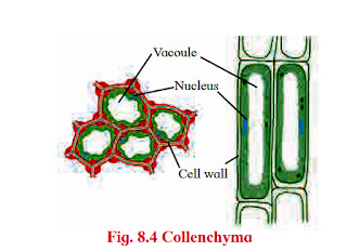
2. Collenchyma :

3. Sclerenchyma :

2. Sclerids

- This is group of cells which have lost the capacity of division and aquired permanent size, shape and functions.
- It is due to different morphological, physiological and functional changes that occur during maturation of the cell.
- Depending upon types of cells, there are two types as -
- Simple permanent tissues and
- Complex permanent tissues.
Simple permanent tissues :

- These are made up of only one type of cells carrying similar functions.
- This tissue is either living or dead.
- Following are the types of simple permanent tissues namely -
- Parenchyma
- Collenchyma and
- Sclerenchyma
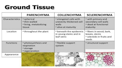
- Cells in this tissue are thin walled, isodiametric, round, oval to polygonal or elongated in shape.
- Cell wall is composed of cellulose.
- Cells are living with prominent nucleus and cytoplasm with large vacuole. This is less specialized permanent tissue.
- Parenchyma has distinct intercellular spaces. Sometimes, cells may show compact arrangement.
- The cytoplasm of adjacent cells is interconnected through plasmodesmata and thus forms a continuous tissue.
- These cells are distributed in all the parts of plant body viz. epidermis, cortex, pericycle, pith, mesophyll cells, endosperm, xylem and phloem.
- These cells store food, water, help in gaseous exchange, increase buoyancy, perform photosynthesis and different functions in plant body.
- Dedifferentiation in parenchyma cells develops vascular cambium and cork cambium at the time of secondary growth.

- It is a simple permanent tissue made up of living cells.
- The cell wall is cellulosic but shows uneven deposition of cellulose and pectin especially at corners.
- The walls may show presence of pits.
- Cells are similar like parenchyma containing cytoplasm, nucleus and vacuoles but small in size and without intercellular gaps. Thus appears to be compactly packed.
- The cells are either circular, oval or angular in transverse section.
- Collenchyma is living mechanical tissue and serves different functions in plants.
- It gives mechanical strength to young stem and parts like petiole of leaf.
- It allows bending and pulling action in plant parts and also prevents tearing of leaf.
- Growth of organs and elongation are other functions.
- Collenchyma is usually absent in monocots and roots of dicot plant.

3. Sclerenchyma :

- It is simple permanent tissue made up of compactly arranged thick walled dead cells.
- The cells are living at the time of production but at maturity they become dead.
- As cells are devoid of cytoplasm their thickened walls are due to uniform deposition of lignin.
- Cells remain interconnected through several pits. It is of two types viz.
- fibres and
- sclerids.
- Fibres are thread-like, elongated and narrow structures with tapering and interlocking end walls.
- These are mostly in bundles, pits are narrow, unbranched and oblique.
- They provide mechanical strength.
2. Sclerids
- Sclerids are usually broad, with blunt end walls.
- These occur singly or in loose groups and their pits are deep branched and straight.
- These are developed due to secondary thickening of parenchyma cells and provide stiffness only.
- This tissue functions as the main mechanical tissue.
- It permits bending, shearing and pulling.
- It gives rigidity to leaves and prevents it from falling. It also gives rigidity to epicarps and seeds.
- Commercial fibres are also produced from sclerenchyma fibres. e.g. jute, flax, hemp.


Part 03 - Complex permanent tissues
Complex permanent tissues :

1. Tracheids :

2. Vessels
Phloem :

1. sieve tubes :
2. sieve cells :

- This tissue is heterogenous comprising of more than one type of cells and all function as a single unit.
- This tissue is involved in conducting the sap and food from source to sink area.
- Xylem and phloem are the complex tissues present in plants.

- It is a dead complex tissue. Components of xylem are -
- tracheids
- vessels
- xylem parenchyma and
- xylem fibres.
- The xylem also provides mechanical strength to the plant body.
- Tracheids and vessels conduct water and minerals. These are also known as hadrome.

- In pteridophytes and gymnosperms tracheids are conducting elements.
- Tracheids are elongated, tubular and dead cells. The ends are oblique and tapering.
- The cell walls are uniformly thickened and lignified. This provides mechanical strength.
- Tracheids contribute 95% of wood in Gymnosperms and 5% in Angiosperms.
- The different types of thickening patterns are seen on their walls such as

- Annular (in the form of rings)
- Spiral (in the form of spring/ helix)
- Scalariorm (ladder like)
- Pitted is most advanced type (small circular area) which may be simple or bordered.
2. Vessels
- Vessels are conducting elements in angiosperms, Selaginella (Pteridophyte) and Gnetum (Gymnosperm) show presence of vessels.
- Vessels are longer than tracheids with perforated or dissolved ends and formed by union of several vessels end to end.
- These are involved in conduction of water and minerals.
- Their lumen is wider than tracheids and the thickening is due to lignin and similar to tracheids.
- In monocots, vessels are rounded where as they are angular in dicot angiosperms.
- The first formed xylem vessels (protoxylem) are small and have either annular or spiral thickenings while latter formed have larger vessels (metaxylem) have reticulate or pitted thickenings.
- When protoxylem is arranged towards pith and metaxylem towards periphery it is called as endarch e.g. in stem and when the position is revert as in the roots is called as exarch.
- Xylem parenchyma cells are small associated with tracheids and vessels. This is the only living tissue among this complex tissue.
- The function is to store food (starch) and sometimes tannins.
- Parenchyma are involved in lateral or radial conduction of water or sap.
- Xylem fibres are sclerenchymatous cells and serve mainly mechanical support. These are called wood fibres.
- These are also elongated, narrow and spindle shaped.
- Cells are tapering at both the ends and their walls are lignified.
Phloem :

- This is a living tissue. It is also called as bast.
- Phloem is responsible for conduction of organic food material from source (leaf generally) to a sink (other plant parts).
- Phloem was named as leptome by Haberlandt as similar to xylem.
- On the basis of origin, it is
- proto (first formed) and
- meta (laterly formed) phloem.


- It is composed of-
- sieve tubes
- sieve cells
- companion cells
- phloem parenchyma and
- phloem fibres.
1. sieve tubes :
- Sieve tubes are long tubular conducting channel of phloem.
- These are placed end to end with bulging at end walls.
- The sieve tube has sieve plate formed by septa with small pores.
- The sieve plates connect protoplast of adjacent sieve tube cells.
2. sieve cells :
- The sieve tube cell is a living cell with a thin layer of cytoplasm but loses its nucleus at maturity.
- The sieve tube cell is connected to companion cell through phloem parenchyma by plasmodesmata.
- Sieve cells are found in lower plants like pteridophytes and gymnosperms.
- The cells are narrow, elongated with tapering ends and sieve area located laterally
- Companion cells are narrow elongated and living.
- These cells are laterally associated with sieve tube elements.
- Companion cells have dense cytoplasm and prominent nucleus.
- Nucleus of companion cell regulates functions of sieve tube cells through simple pits.
- From origin point of view, sieve tube cells and companion cell are derived from same cell.
- Death of the one results in death of the other type.
- Phloem parenchyma cells are living, elongated found associated with sieve tube and companion cells.
- The chief function is to store food, latex, resins, mucilage, etc.
- The cells carry out lateral conduction of food material.
- These cells are absent in most of the monocots.
- Phloem fibres are the only dead tissue among this unit.
- These are sclerenchymatous.
- Generally absent in primary phloem, but present in secondary phloem.
- These cells are with lignified walls and provide mechanical support.
- These are used in making ropes and rough clothes.

Part 04 - Tissue Systems
Tissue Systems :

- Plant tissues are derived from meristems and their structure and functions depend on the position.
- Types of tissue systems [ On the basis of their structure and location] :
- Epidermal tissue system
- ground tissue system and
- vascular tissue system.

- It forms the outer covering of plant body and is derived from protoderm or dermatogen.
- The two types of structures are seen in epidermal tissue system viz
- epidermis and
- epidermal appendages.
- Epidermis is the outermost protective cell layer made up of compactly arranged cells without intercellular spaces.
- Cells show presence of central large vacuole, thin cyctoplasm and a nucleus.
- The outer side of the epidermis is often covered with a waxy thick layer called the cuticle which prevents the loss of water.
- It may bear hairs. Root epidermis has root hairs.
- These are unicellular elongated and involved in absorption of sap from the soil.


- In stem, epidermal hairs are called trichomes.
- These are generally multicellular, branched or unbranched, stiff or soft or even secretory.
- These help in preventing water loss due to transpiration.
- Small gateways in the epidermal cells are called as stoma. Such stoma are controlled or guarded by specially modified cells called guard cells.
- These guard cells may be kidney shaped (dicot) or dumbbell shaped (monocot), collectively called as Stomata.
- Stoma, guard cells and subsidiary cells form a unit called stomatal apparatus.
- Stomata are further covered by subsidiary cells.
- Guard cells have chloroplasts to carry out photosynthesis.
- Guard cells change their turgor pressure causing its opening and closing, thus they play a vital role in exchange of gases and water vapour.
- All the plant tissues excluding epidermal and vascular tissue is ground tissue.
- It is made up of simple permanent tissue e.g. paranchyma.
- It is present in cortex, pericycle, pith and medullary rays in the primary stem and root.
- Collenchyma and schlerenchyma in the hypodermis and chloroplasts containing mesophyll tissue in leaves is also ground tissue.

- These are the distinct patches of the complex tissue viz. Xylem and phloem.
- On the basis of their arrangement in the plant body these are radial when both the complex tissue are situated separately on separate radius as separate bundle. This is a common feature of roots.
- In the stem, the complex tissue is collectively present as neighbours of each other on the same radius in the form of xylem inside and phloem outside hence called conointcollateral vascular bundles.
- These bundles may be further of open type (secondary growth takes place) containing cambium in between them and closed type if cambium is not present (secondary growth absent).
- When phloem is present in a vascular bundle on both the sides of xylem and intervening cambium tissue, it is called bicollateral vascular bundle.
- It is a feature of family Cucurbitaceae.
- When one vascular tissue is completely encircling the other, it is called as concentric vascular bundle, this may be leptocentric (phloem encircled by xylem) or hadrocentric (xylem encircled by phloem).
- When one complex tissue is encircling on both the faces of the other it is amphicribral (xylem encircled by phloem on both faces) and amphivasal (phloem encircled by xylem on both faces).
- The vertical growth of the roots and stems in length with the help of apical meristem is called as primary growth.
- Dicotyledonous plants and gymnosperms exhibit increase in girth of root and stem.
- In dicot stem, secondary growth begins with the formation of a continuous cambium ring.
- The cambium present between the primary xylem and primary phloem of a vascular bundle is called intraasicular cambium.
- The cells of medullary rays adjoining these intrafascicular cambium strips become meristematic (regain the capacity to divide) and form the interascicular cambium.
- Thus a complete and continous ring of vascular cambium is formed.
- The cambium ring cuts off new cells, towards both the sides, inner and outer.
- The cells that are cut-off towards pith (inner side) mature into secondary xylem and cells that are cut-off towards periphery mature into secondary phloem.
- Generally, amount of secondary xylem is more than the secondary phloem.
- In woody plants, secondary tissues constitute the bulk of the body. This provides support, conduction of water and minerals and protection.
- Lateral meristems play a major role in development of secondary tissues.

- With the onset of favourable season, meristematic cells of intrafascicular cambium become active.
- Simultaneously, the ray parenchyma cells, both fusiform initials and ray initials become meristematic. This is known as dedierentiation.
- These form patch of cambial cells (meristematic cells) in between the adjacent bundles and produce interfascicular cambium.
- Now both intrafascicular and interfascicular cambium join and form a complete ring. This is known as cambial ring.
- This is possible because they lie in one plane.
Secondary growth in roots :
- It is also observed in most of the dicot and gymnospermic roots by producing secondary vascular tissue and periderm.
- Secondary growth is produced by vascular cambium and cork cambium respectively.
- Conjuctive parenchyma cells present on the inner edges of primary phloem bundles become meristematic.
- These cells add secondary xylem and secondary phloem on the inner and outer side respectively.
- These events are similar to secondary growth in stems.
Part 05 - Wood
Wood :
- During favourable conditions, spring ood (early wood) is formed which has broader xylem bands, lighter colour, tracheids with thin wall and wide lumen, fibres are less in number, low density.
- Whereas, during unfavourable season autumn ood (late wood) is formed which has narrow xylem band, darker in colour, lumen is narrow and walls are thick with abundant fibres are present of high density.
- Tracheary elements of heartwood are plugged by in-growth of adjacent parenchyma cells i.e. tyloses.
- They are filled by oils, gums, resins, tannins called as extractives.
- Thus inner non-functinal, durable part which is resistant to pathogens is called duramen or heartwood.
- Outer light, functional part of secondary xylem, cells are living, no deposition, lighter and less durable, more susceptible to pathogens and involved in conduction of sap is called as sap ood (alburnum).
Part 06 - Cork cambium and secondary growth
Cork cambium and secondary growth:
- Increase in diameter of stem by secondary growth is mainly due to the activity of vascular cambium present the outer cortical layer.
- When epidermis gets ruptured, it becomes necessary to replace these cells by new cells. Phellogen (cork cambium) develops in extrastelar region of stem.
- The outer cortical cells of cortex become meristematic and produce a layer of thin walled, rectangular cells.
- These cells cut off new cells on both sides.
- The cells produced on outer side develop phellem (cork) wheras on the inner side produce phelloderm (secondary cortex).
- The cork is impervious in nature and does not allow entry of water due to suberized walls. Secondary cortex is parenchymatous in nature.
- Phellogen, phellem and phelloderm constitute periderm.
- Activity of cork cambium develops a pressure on the other cells and these cells die.
- Bark is non-technical term refering to all cell types found external to vascular cambium including secondary phloem.
- Bark of early season is soft and of the late season is hard.
- Lenticles are aerating pores present as (raised scars) the surface of bark.
- These are portions of periderm, where phellogen activity is more, lenticles are means for gaseous and water vapour exchange.
- Monocot stems lack cambium hence secondary growth does not take place.
- But accessory cambium development in plants like, Dracena, Agave, Palms and root of sweet potato show presence of secondary growth. This is called as anomalous secondary growth
Part 07 - Anatomy of Root, Stem and Leaf
Anatomy of Root, Stem and Leaf :
A. Anatomy of Dicot Root :


A. Anatomy of Dicot Root :

- The transverse section of a typical dicotyledonous root shows following anatomical features.
- The outermost single layer of cells without cuticle is Epiblema.
- Some of its cells are prolonged into unicellular root hair.
- Next to it is the Cortex which consists of several layers of typical parenchymatous cells.
- After the death of epiblema, outer layer of cortex become cutinized and is called Exodermis.
- The cortical cells store food and water. The innermost layer of cortex is called Endodermis.
- The cells are barrel-shaped and their radial walls bear Casparian strip or Casparian bands composed of suberin.
- Near the protoxylem, there are unthickened passage cells.
- A single layer of parenchymatous Pericycle is present just below endodermis which bounds the stele or vascular cylinder.
- Stele consists of 2 to 6 radial vascular bundles.
- Xylem is exarch.
- Based on the number of groups of xylem and phloem, the stele may be diarch to hexarch.
- A parenchymatous connective tissue or conjunction tissue is present between xylem and phloem.
- The central part of stele or vascular cylinder is called Pith.
- It is narrow and made up of parenchymatous cells, with or without intercellular spaces.
- At later stage, a cambium ring develops between xylem and phloem which causes secondary growth in thickness.
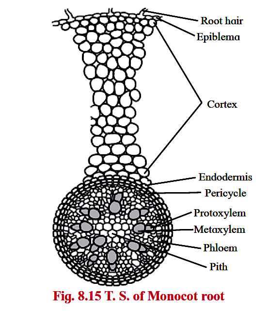
- It resembles that of a dicot root in its basic plan.
- However, it possesses more than six xylem bundles (polyarch condition).
- Pith is large and well-developed.
- Secondary growth is absent.
Part 08 - Anatomy of Dicot Stem (Sunflower)
Anatomy of Dicot Stem (Sunflower) :
A transverse section of dicot stem shows the following structures :
 1. Hypodermis :
D. Anatomy of Monocot Stem :

A transverse section of dicot stem shows the following structures :
- Epidermis is single, outermost layer with multicellular outgrowth called trichomes.
- A layer of cuticle is usually present towards the outer surface of epidermis.
- Cortex is situated below the epidermis and is usually differentiated into three regions namely-
- hypodermis
- general cortex and
- endodermis

- Hypodermis is situated just below the epidermis and is made of 3-5 layers of collenchymatous cells.
- Intercellular spaces are absent.
- General cortex is made up of several layers of large parenchymatous cells with intercellular spaces.
- Endodermis is an innermost layer of cortex which is made up of barrel shaped cells. It is also called starch sheath.
- Stele is the central core of tissues differentiated into pericycle, vascular bundles and pith.
- Pericycle is the outermost layer of vascular system situated between the endodermis and vascular bundles.
- In sunflower, it is multilayered and also called hard bast.
- Vascular bundles are conjoint, collateral, open, and are arranged in a ring.
- Each one is composed of xylem, phloem and cambium.
- Xylem is endarch.
- A strip of cambium is present between xylem and phloem.
- Pith is situated in the center of the young stem and is made up of large-sized parenchymatous cells with conspicuous intercellular spaces.

- It differs from dicot.
- Epidermis is without trichomes and the hypodermis is sclerenchymatous.
- Vascular bundles are numerous and are scattered in ground tissue.
- Each vascular bundle is surrounded by a sclerenchymatous bundle sheath.
- Vascular bundles are conjoint, collateral and closed (without cambium).
- Xylem is endarch and shows lysigenous cavity.
- Pith is absent.
- Secondary growth is also absent.
Part 09 -Anatomy of Leaf
Anatomy of Leaf :
V. S. of Typical dicot leaf :
 Palisade parenchyma
Isobilateral Leaf :

- Dorsiventral Leaf is very common in dicotyledonous plants where the mesophyll tissue is differentiated into
- palisade and
- spongy parenchyma.
- The leaves are commonly horizontal in orientation with distinct upper and lower surfaces.
- The upper surface which faces the sun is darker than the lower surface.
V. S. of Typical dicot leaf :
- Upper epidermis consists of a single layer of tightly packed rectangular, barrel shaped, parenchymatous cells which are devoid of chloroplast.
- A distinct layer of cuticle lies on the outside of the epidermis.
- Stomata are generally absent.
- Between upper and lower epidermis, there is chloroplast-containing photosynthetic tissue called Mesophyll.
- Mesophyll is differentiated into -
- palisade tissue and
- spongy tissue.

- It is present below upper epidermis and consists of closely packed elongated cells.
- The cells contain abundant chloroplasts and help in photosynthesis.
- It is present below palisade tissue and consists of loosely arranged irregularly shaped cells with intercellular spaces.
- The spongy parenchyma cells contain chloroplast and are in contact with atmosphere through stomata.
- Vascular system is made up of a number of vascular bundles of varying size depending upon the venation.
- Each one is surrounded by a thin layer of parenchymatous cells called bundle sheath.
- Vascular bundles are closed and xylem towards upper epidermis and phloem towards lower epidermis.
- Cambium is absent hence no secondary growth in the leaf.
- Lower epidermis consists of a single layer of compactly arranged rectangular, parenchymatous cells.
- A thin layer of cuticle is also present.
- The lower epidermis contains a large number of microscopic pores called stomata.
- There is an air-space called substomatal chamber at each stoma.
Isobilateral Leaf :
- In this leaf both the surfaces are equally illuminated as both the surface can face the sun, and show similar structure.
- The two surfaces are equally green.
- Generally monocotyledonous plants have isobilateral leaves.

- resembles a dicot leaf in its anatomical structure.
- However, it shows stomata on both the surfaces and mesophyll is not differentiated into palisade and spongy tissue.
- It has parallel veins.
- These are conjoint, collateral and closed.
Source From Internet
No comments:
Post a Comment